Home » Without Label » Arteries Diagram - Semara's Mystifying Anatomy: The Veins and Arteries Below ... - The heart receives its own supply of blood from the coronary arteries.
Arteries Diagram - Semara's Mystifying Anatomy: The Veins and Arteries Below ... - The heart receives its own supply of blood from the coronary arteries.
Arteries Diagram - Semara's Mystifying Anatomy: The Veins and Arteries Below ... - The heart receives its own supply of blood from the coronary arteries.. A condition which arises spontaneously or as the result of trauma, where the walls of the artery are split, leading to internal bleeding and disruption of blood flow. Arteries and veins are two of the body's main type of blood vessels. After receiving blood directly from the left ventricle of the heart, the. Finally, the smallest arteries, called arterioles are further branched into small capillaries, where the exchange of all the nutrients, gases and other waste molecules are carried out. Though more often occurring with carotid arteries (the other major ones supplying the brain through the neck), vertebral arteries can be impacted.
This is an excellent human heart diagram which uses different colors to show different parts and also labels a number… Proximal structures of the upper extremity 9p image quiz. Constricted arteries oppose blood flow, and more pressure is required to push blood. Inner body parts with their names. Transposition of the great arteries is a serious but rare heart defect present at birth (congenital), in which the two main arteries leaving the heart are reversed (transposed).
Foods To Help Prevent Clogged Arteries! from www.shakahariblog.com Arteries of the body diagram. Ascending aorta, aortic arch, thoracic aorta, and abdominal aorta. Arteries carry blood away from the heart in two distinct pathways: Transposition of the great arteries is a serious but rare heart defect present at birth (congenital), in which the two main arteries leaving the heart are reversed (transposed). Arteries and arterioles carry oxygenated blood _____ from the heart to the body. Anatomy and function of the coronary arteries. Each of these arteries delivers blood to the leg and continues into the foot, with the posterior tibial and fibular arteries forming the plantar arteries and plantar arch that supply blood to the bottom of the foot and toes. The veins also lack the elastic internal lamina that lies.
The arteries of the brain.
This is a list of arteries of the human body. Spinal cord and meninges 12p image quiz. Create healthcare diagrams like this example called arteries of the foot in minutes with smartdraw. These arteries and their branches supply all parts of the heart muscle with blood. A condition which arises spontaneously or as the result of trauma, where the walls of the artery are split, leading to internal bleeding and disruption of blood flow. Arteries are components of the cardiovascular system. Arteries and veins are two of the body's main type of blood vessels. Smartdraw includes 1000s of professional healthcare and anatomy chart templates that you can modify and make your own. Arteries are blood vessels that carry blood away from the heart. Ascending aorta, aortic arch, thoracic aorta, and abdominal aorta. Arteries of the body diagram. The triangles of the neck. The aorta branches into a network of smaller arteries that extend throughout the body.
Blood carried by arteries is usually highly oxygenated, having just left the lungs on its way to the body's tissues. Each artery is a muscular tube lined by smooth tissue and has three layers: Start studying review of arteries. The two exceptions are the pulmonary and the umbilical arteries, which carry deoxygenated blood to the organs that oxygenate it (lungs and placenta. The arteries of the brain.
CIRCULATORY SYSTEMS from www2.estrellamountain.edu Anatomy and function of the coronary arteries. Smartdraw includes 1000s of professional healthcare and anatomy chart templates that you can modify and make your own. A probe placed against the skin reflects sound waves off the carotid artery, and a computer constructs images on a screen.doppler ultrasound can be. Abdomen arteries, veins, and duct diagram. The two exceptions are the pulmonary and the umbilical arteries, which carry deoxygenated blood to the organs that oxygenate it (lungs and placenta. The veins also lack the elastic internal lamina that lies. Each of these arteries delivers blood to the leg and continues into the foot, with the posterior tibial and fibular arteries forming the plantar arteries and plantar arch that supply blood to the bottom of the foot and toes. The tunica medica, which is the very muscular middle layer in arteries, is thinner and less muscular in veins.
Transposition of the great arteries is a serious but rare heart defect present at birth (congenital), in which the two main arteries leaving the heart are reversed (transposed).
Resistance (r) the force opposing blood flow. Inner body parts with their names. This is a congenital defect in which the left pulmonary artery branches off the right pulmonary artery, rather than directly from the pulmonary trunk. More artery diagrams are posted in the following 101 diagramss below. Learning all of the major arteries and veins of the cardiovascular system at once is a pretty large undertaking, so in the following sections, we're going to focus on the arteries of the cardiovascular system. Anatomy and function of the coronary arteries. Constricted arteries oppose blood flow, and more pressure is required to push blood. After receiving blood directly from the left ventricle of the heart, the. Arteries are components of the cardiovascular system. Veins are the blood vessels present throughout the body. In this image, you will find right gastric artery, common hepatic artery, celiac trunk, left gastric artery, splenic artery, splenic vein, pancreas, suprarenal vein, renal vein, renal artery, inferior mesenteric vein , gonadal vein, gonadal artery, two alternative position of artery, left colic artery. The tunica medica, which is the very muscular middle layer in arteries, is thinner and less muscular in veins. Transposition of the great arteries is a serious but rare heart defect present at birth (congenital), in which the two main arteries leaving the heart are reversed (transposed).
The arteries' smaller branches are called arterioles and capillaries. The subclavian artery runs into the axillary region where it becomes known as the axillary artery. Though more often occurring with carotid arteries (the other major ones supplying the brain through the neck), vertebral arteries can be impacted. An artery is an elastic blood vessel that transports blood away from the heart. In this image, you will find right gastric artery, common hepatic artery, celiac trunk, left gastric artery, splenic artery, splenic vein, pancreas, suprarenal vein, renal vein, renal artery, inferior mesenteric vein , gonadal vein, gonadal artery, two alternative position of artery, left colic artery.
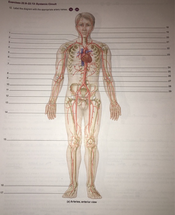
Solved: Label The Diagram With The Appropriate Artery ... from media.cheggcdn.com A condition which arises spontaneously or as the result of trauma, where the walls of the artery are split, leading to internal bleeding and disruption of blood flow. Create healthcare diagrams like this example called arteries of the foot in minutes with smartdraw. Smartdraw includes 1000s of professional healthcare and anatomy chart templates that you can modify and make your own. The typical configuration consists of two coronary arteries, a left main coronary artery (lmca) and a right coronary artery (rca), arising from the left posterior and right anterior aortic or coronary sinuses respectively, in the proximal ascending aorta.these are the only two branches of the ascending aorta. Ear anatomy 17p image quiz. The aorta branches into a network of smaller arteries that extend throughout the body. This is the opposite function of veins, which transport blood to the heart. Veins are the blood vessels present throughout the body.
Start studying review of arteries.
Arteries of the head and neck diagram art print vintage anatomy art print on tea stained paper dog art dog s wfh office art. Create healthcare diagrams like this example called arteries of the foot in minutes with smartdraw. Resistance (r) the force opposing blood flow. Abdomen arteries, veins, and duct diagram. Ear anatomy 17p image quiz. Veins are the blood vessels present throughout the body. Ascending aorta, aortic arch, thoracic aorta, and abdominal aorta. The arteries of the upper extremity. An artery (plural arteries) (from greek ἀρτηρία (artēríā) 'windpipe, artery') is a blood vessel that takes blood away from the heart to one or more parts of the body (tissues, lungs, brain etc.). The triangles of the neck. Posterior view of the left lower extremity 6p image quiz. The typical configuration consists of two coronary arteries, a left main coronary artery (lmca) and a right coronary artery (rca), arising from the left posterior and right anterior aortic or coronary sinuses respectively, in the proximal ascending aorta.these are the only two branches of the ascending aorta. Let's now see how we can revise them with the help of cardiovascular system diagram activities.